لا توجد دقة أعلى متوفرة.
PET-image.jpg (373 × 405 بكسل، حجم الملف: 48 كيلوبايت، نوع الملف: image/jpeg)
Summary
| Description |
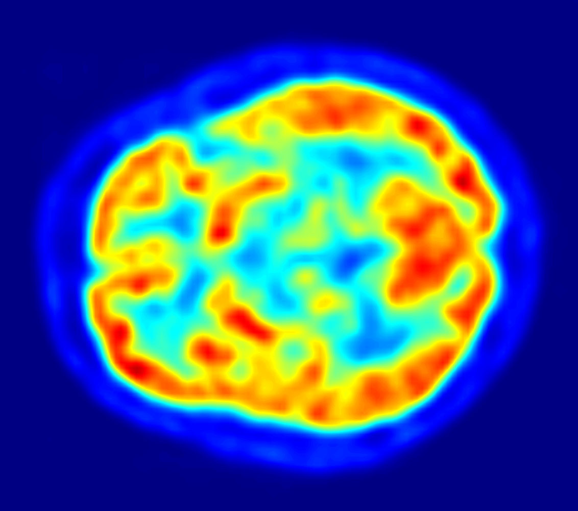
This is an image taken from a typical PET acquisition. It is a tomographic view of a brain examination in transaxial view. Red areas show more accumulated radioactivity and blue areas are partions where low to no activity was accumulated. It should illustrate how a typical PET image looks like. It was taken with an ECAT Exact HR+ PET Scanner. |
||
|---|---|---|---|
| Source |
own work - part of master thesis |
||
| Date |
April 2003 |
||
| Author |
Jens Langner (http://www.jens-langner.de/) |
||
| Permission (Reusing this image) |
|
وصلات
الصفحات التالية تحتوي على وصلة لهذه الصورة:
بيانات ميتا
هذا الملف يحتوي معلومات إضافية، غالبا ما تكون أضيفت من قبل آلة التصوير الإلكترونية أو الماسح الضوئي المستخدم في تحميل الصورة إلى الحاسوب. إذا كان الملف قد عُدّل عما كان عليه عند تحميل الصورة فإن المعلومات الواردة هنا قد لا تعبر عن هذه الصورة المعدلة.
| التوجيه | وضعية طبيعية |
|---|---|
| الدقة الأفقية | 72 نقطة لكل بوصة |
| الدقة الرأسية | 72 نقطة لكل بوصة |
| البرمجيات المستخدمة | QuickTime 7.0.1 |
| تاريخ و وقت تغيير الملف | 15:32، 14 يونيو 2005 |
| وضع Y و C | 1 |