حجم هذا العرض: 422 × 600 بكسل
الصورة بدقة كاملة (795 × 1,130 بكسل، حجم الملف: 290 كيلوبايت، نوع الملف: image/png)
This image was copied from wikipedia:pt. The original description was:
- Descrição:
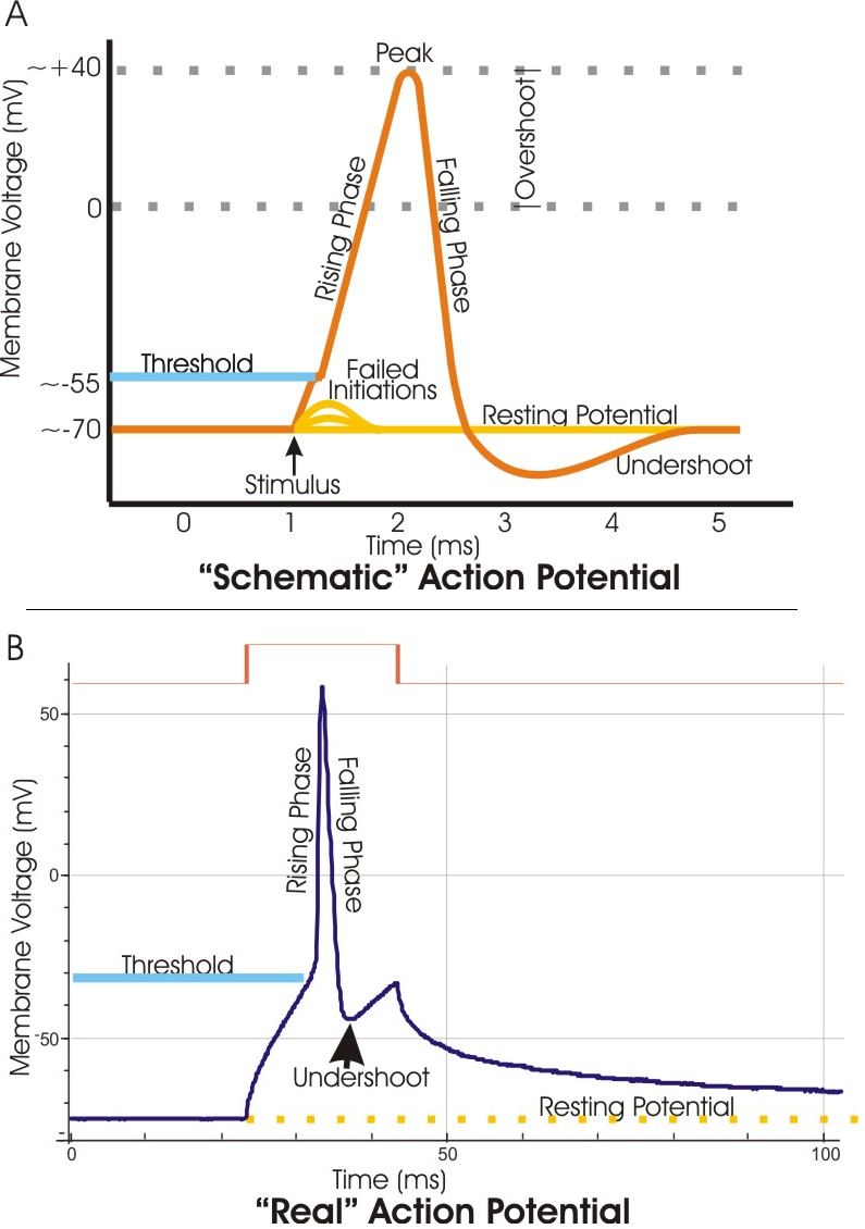
Representação esquemático de um potencial deacção.
- Fonte e créditos:
- Licença:
 |
Permission is granted to copy, distribute and/or modify this document under the terms of the GNU Free Documentation license, Version 1.2 or any later version published by the Free Software Foundation; with no Invariant Sections, no Front-Cover Texts, and no Back-Cover Texts. A copy of the license is included in the section entitled "GNU Free Documentation license".
| | | | | | | | | | | | | | | | | | | | | | | | | | | | Ido | | | | | | | | | | | | | | | | | | | | | | | | | | | | | | | | | | | | +/- |
| date/time | username | resolution | size | edit summary |
|---|---|---|---|---|
| 02:54, 27 Julho 2006 | Vulpes | 795×1130 | 297 |
Description from other copy (Image:422px-Action potential vert.png):
from en:Image:Action potential vert.png
Modified version of older Image:Action potential reloaded.jpg.
Caption (from action potential):
A. Schematic of an electrophysiological recording of an action potential showing the various phases which occur as the wave passes a point on a cell membrane. B. An actual action potential (blue trace) recorded from a mouse hippocampal pyramidal neuron. In this case, the action potential was stimulated by a prolonged pulse of current (brown trace; approx. 2 micro Amps)passed into the cell through the recording electrode. This method of stimulation distorts the AP compared to the schematic, in that the "real" action potential is sitting atop a voltage offset caused by the current pulse. Thus, for example, the "undershoot" is offset from the resting potential, although it would dip below rest if the offset were not present. The slow decline of the membrane potential back toward rest upon the termination of the current pulse reflects the long time constant of the neuronal membrane.
وصلات
الصفحات التالية تحتوي على وصلة لهذه الصورة: